
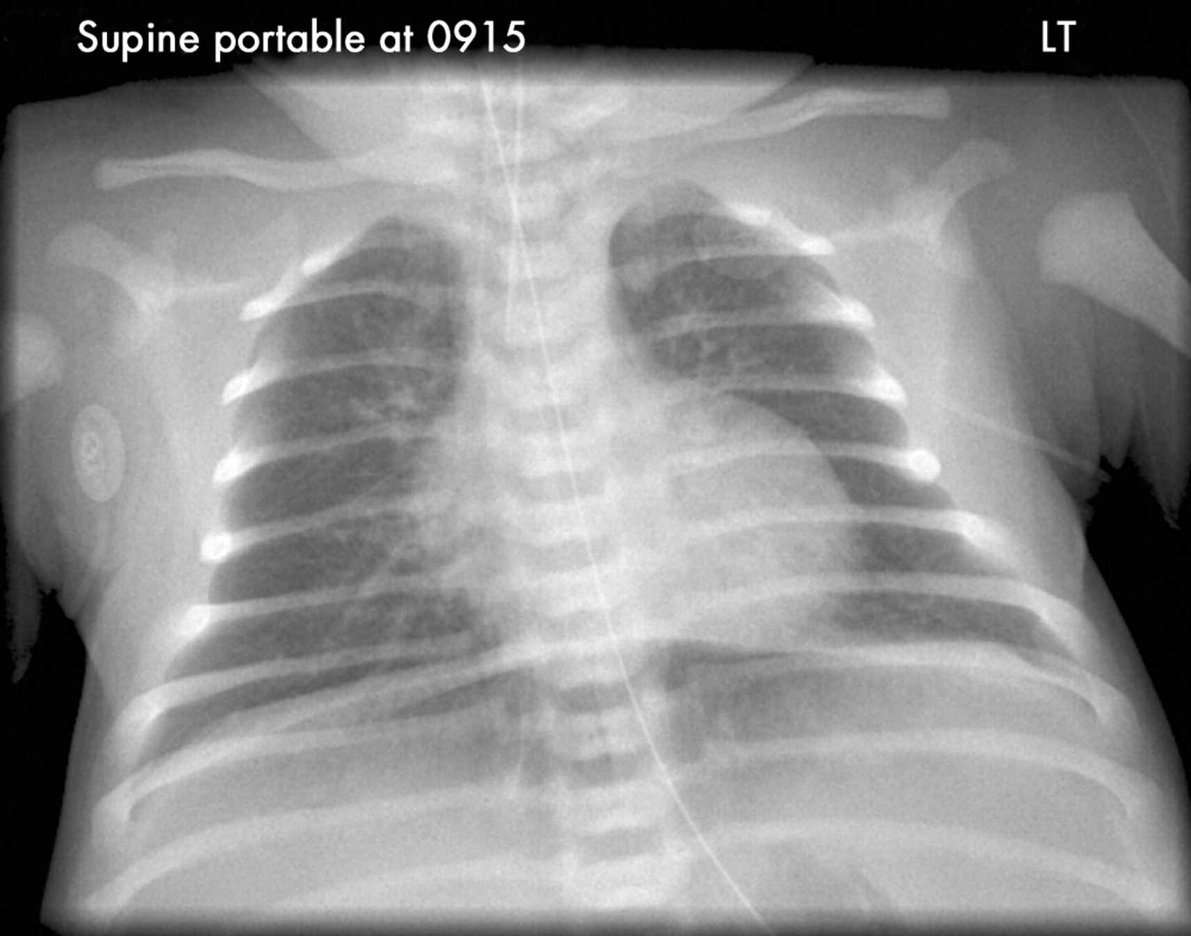
The distal end of the drainage tube must remain under the water surface levelThere is always an outlet to the atmosphere to allow air to escapeIt is suitable for use with a simple pneumothorax when the vent is left open to the atmosphere or following a. The gastric air bubble on the left.

Chest Xray Film Of A Posttraumatic Patient Showing Free Air Under Dome Of Right Diaphragm From Hollow Abdominal Organs Rupture Stock Photo Picture And Royalty Free Image Image 109400946
![]()
Chest X Ray Erect View Shows Air Under Diaphragm Marked By Arrow Download Scientific Diagram

False Appearance Of Free Air Under The Diaphragm The American Journal Of The Medical Sciences
It has 3 major characteristics that can be found on chest X-Ray.

Air under diaphragm cxr. Clinical cases and self assessment to enhance interpretation skills through various Chest X-Ray problems. C CXR in a different patient shows multiple ill-defined reticulo-nodular lesions in both lungs with basal predominance suggestive of miliary TB. Look for free air under diaphragm on upright projections.
Trauma is the third leading cause of death in all age groups after cardiovascular diseases and cancer1 However trauma is the most common cause of death in the age of first four decades2 Although trauma-related injuries can occur in many parts of body one out of four trauma patients die due to thoracic injury or its complications3. Right hemidiaphragm Should be higher than the left. FIGURE 76-6 There is cardiac enlargement splaying of the carina indicating left atrial enlargement prominent pulmonary vasculature and hazy opacification centrally suggestive of a left-to-right shunt at PDA level.
Atelectasis describes small areas of collapsed lung. B CXR in a different patient shows multiple coalescent air-space nodules in RT upper zone. JSS Medical College Mysuru THORACO-ABDOMINAL SIGN A sharply marginated mediastinal mass projected over the diaphragm on a CXR or on an AXR will lie wholly or partly in the thoraxbecause it is outlined by the air in the lung.
Chest decompression in the field Needle decompression Finger thoracostomy Needle decompression is performed preferably at the 5 th intercostal space mid axillary line with the arm in the abducted position 3This location may not be easily accessible and in such circumstances the 2 nd intercostal space mid axillary line is used. It is often supplemented by an upright PA view of the chest to rule out air under the diaphragm or thoracic etiologies presenting as abdominal complaints and a standing view of the abdomen to differentiate obstruction from ileus by examining gastrointestinal airwater levels. Atelectasis and collapse both describe the same pathophysiology though atelectasis tends to be used to describe small areas of lung that are not fully expanded whereas collapse tends to be used to describe larger more confluent areas.
Pleuritic chest pain is characterized by sudden and intense sharp stabbing or burning pain in the chest when inhaling and exhaling. Example of a Complete History and Physical Write-up Patient Name. The dome of the diaphragm should project at the level of the 8th10th posterior ribs if the mean airway pressure is appropriately adjusted.
Imaging if concerned for acute abdomen must include chest X-ray to rule out perforation as demonstrated by free air under the diaphragm. LITFL Top 100 CXR quiz. Patient who is reliable and old CPMC chart.
Like all methods of radiography chest radiography employs ionizing radiation in the form of X-rays to generate images of the chest. Postoperative pulmonary complications PPCs are common costly and increase patient mortality. The most obvious finding on this CXR is free air under the diaphragm.
This is a basic article for medical students and other non-radiologists. If much higher think of effusion lobar collapse diaphragmatic paralysis. Reveal cause Urgent laparotomy repair Ruptured AAA Elderly Severe generalised abdominal pain Back pain Reduced GScollapse Shock Peritonitis Expansile mass USS abdomen if freely available CT only.
A CXR depicts RT upper zone consolidation with prominent RT hilum. CXR was without pleural effusion or free air under the diaphragm. Academiaedu is a platform for academics to share research papers.
CXR flat and upright perforated lumen with free. 1 bottle The simplest form of underwater seal drainage systems. In patients who are upright when imaged such as for a PAlateral CXR air in the pleural space from a pneumothorax tends to collect in non-dependent locations such as.
Here are Jan 11 2020 Hiatal hernia is a condition where a portion of the stomach pushes into the chest through the hiatus an opening in the diaphragm. The patient rinsed out his mouth with water. Chest radiographs are the most common film taken in medicine.
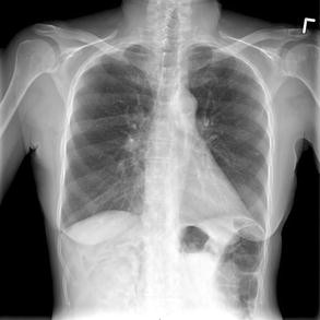
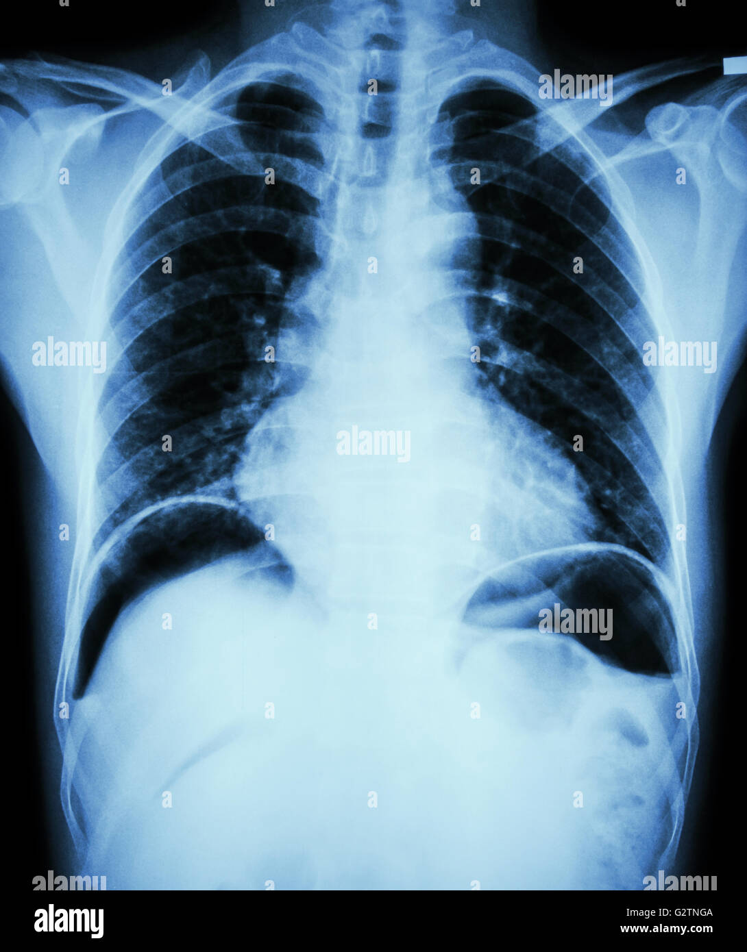
Right hemidiaphragm should be higher than the left Figure-11. Perforation of the bowel is seen as free air under the diaphragm on a CXR. The outline of the diaphragm should be clear and smooth.
GI performed an urgent endoscopy which demonstrated Grade 2a lesions. D Diaphragm. If air is found in this location its called pneumoperitoneum which is a medical emergency.
Pulmonary embolism is the most common serious cause found in. A chest radiograph called a chest X-ray CXR or chest film is a projection radiograph of the chest used to diagnose conditions affecting the chest its contents and nearby structures. Air should never appear in the peritoneal cavity outside the gastrointestinal tract.
This system can drain both fluid and air. He developed drooling and chest pain. Air under diaphragm CT abdopelvis.
Free Air Under Diaphragm Yellow. Changes to the respiratory system occur immediately on induction of general anaesthesia. This finding indicates a bowel perforation unless when the patient had recent abdominal surgery and there is still some air left in the abdomen which can stay there for several days.
This is the 3rd CPMC admission for this 83 year old woman with a long history of hypertension who presented with the chief complaint of substernal toothache like chest pain of 12 hours. If you cannot see parts of the diaphragm consider infiltrate or effusion. Lab work was obtained which demonstrated an anion-gap acidosis likely due to the sulphur ingestion.
If film is taken in erect or upright position you may see free air under the diaphragm if intra-abdominal perforation is. Ribs sternum spine clavicles symmetry fractures dislocations lytic lesions density Soft tissues looking for symmetry swelling loss of tissue planes subcutaneous air masses. Chest radiographs in active TB.
The diaphragmatic contour looks like a dome shape and the right side located little higher than the. Anyone who experiences bibasilar crackles and shortness of breath chest pain or blood-tinged mucus should seek immediate medical attention. The diaphragm should be indistinguishable from the underlying liver in healthy individuals on an erect chest X-ray however if free gas is present often as a result of bowel perforation air accumulates under the diaphragm causing it to lift and become visibly separate from the liver.
A KUB is a plain frontal supine radiograph of the abdomen. Respiratory drive and muscle function are altered lung volumes reduced and atelectasis develops in 75 of patients receiving a neuromuscular blocking drug. In CXR interpretation it is common to leave soft tissues until the end.
It can be caused by a ruptured appendix perforated ulcer or ruptured diverticulum. A large bore IV Cannula 14 or 16G with a 10ml syringe.
Perforation How To Spot Free Intraperitoneal Air On Abdominal Radiograph Adc Education Practice Edition

Air Under Diaphragm Chest Xray Erect Stock Photo Edit Now 1064432564
Plain Chest X Ray Revealed Free Air Under The Right Diaphragm Black Download Scientific Diagram
Rigler Sign A Subtle Finding Of Pneumoperitoneum Springerlink

Subdiaphragmatic Free Gas Radiology Reference Article Radiopaedia Org
Pneumoperitoneum Wikiradiography

Chest X Ray Pneumoperitonuem Air Under Diaphragms Youtube
Peptic Ulcer Perforate Film Chest X Ray Show Free Air Under Dome Of Both Diaphragm Due To Air Leak From Gastric Ulcer Or Duo Stock Photo Alamy


