
However healing times vary depending on which bone is broken. Patella fracture - X-ray appearances.

X Ray Of Rheumatoid Arthritis In The Feet
Abnormal
3 871 Foot Xray Stock Photos Pictures Royalty Free Images Istock

Foot and Ankle Arthritis Types.
Normal foot x ray. We found that emergency doctors were ordering many imaging studies for ankle injuries that were then found to be normal. Plain X-ray needs to be included in all trauma and overuse presentations. Craniosynostosis an abnormal fusion of two or more cranial bones.
Rarely does a patient need both an ankle and foot x. X-Ray is limited to examining a few body conditions only. J Ultrasound Med 200019315-21.
B X-Ray of your foot. Intestinal ultrasonography in children and young adults. You dont need to jump straight to an MRI.
An X-ray can usually do a great job if there is a foreign object or if there is an extremely large bone prominence. Welcome To Northwest Foot Ankle PS. Synostoses is fusion of two or more bones.
Search for magnetic resonance for physics details. Many stress fractures of the foot or ankle will heel in 4 to 6 weeks. Sutter Davis Hospital is a technologically advanced acute care hospital offering care for residents in Davis Dixon Winters Woodland West Sacramento Vacaville and rural communities throughout Yolo and Eastern Solano counties.
As a general rule the clustering of small bones in this area can make the interpretation of plain X-ray more challenging. That is why we offer the highest standard of care in podiatry. Acute traumatic injuries are treated with splinting where chonic injuries often require surgical reconstruction.
Learning radiology of knee injury covering fractures of the tibia and patella - Lower limb X-rays - Knee fractures as seen on X-ray Fractures of the tibial plateau. Lateral chest X-ray. Shoe-fitting fluoroscopes also sold under the names X-ray Shoe Fitter Pedoscope and Foot-o-scope were X-ray fluoroscope machines installed in shoe stores from the 1920s until about the 1970s in the United States Canada United Kingdom Australia South Africa Germany and Switzerland.
Knee joint effusion with haemarthrosis. When checking any post-traumatic foot X-ray it is crucial to assess alignment of the bones at the joints. Pre-contrast T1 weighted sagittal.
Wiersma F Sramek A Holscher HC. On a chest x-ray lung abnormalities will either present as areas of increased density or as areas of decreased density. Chest x-ray review is a key competency for medical students junior doctors and other allied health professionals.
A bunion is a bony hump that forms at the base of the big toe where it attaches to the foot. The radiation is created when an electric current is generated from a high voltage generator causing electrons to boil-off from the cathode end of an X-ray tube assembly. When considering conditions being screened for with imaging there is a lower threshold for using further imaging modalities in mid-foot assessment.
MRI is more versatile than the X-Ray and is used to examine a large variety of medical conditions. Healing time varies so ask your doctor when you can resume normal activities. An X-ray can usually be better than advanced imaging for bone assessment.
In the UK they were known as Pedoscopes after the company based in St. The chest radiograph also known as the chest x-ray or CXR is anecdotally thought to be the most frequently-performed radiological investigation globally although no published data is known to corroborate thisUK government statistical data from the NHS in England and Wales shows that the chest radiograph remains consistently the most frequently requested imaging test by GPs 2019 dataset 5. The technologist plays a pivotal role in improving diagnostic accuracy by providing diagnostic images1 This requires a technologist to be aware of the various.
The Ottawa Ankle Rule rules out clinically significant foot and ankle fractures to reduce use of x-ray imaging. Radioulnar synostosis the abnormal fusion of the radius and ulna. Often the big toe deviates toward the second toe.
Our Foot Doctor and Foot Surgeon provides expert care diagnosis and treatment of ankle and. Find relief from foot ankle pain with top rated Podiatrist Shawn Echard of the Laurel Podiatry and The Foot and Ankle Laser Institute located in Greensburg Somerset Mount Pleasant and Pittsburgh PA. Photo courtesy of Evelyn Taylor.
As you get older you have a higher risk of arthritisThe joint damage from this condition can cause swelling pain and physical changes in your feet and ankles. Radiology Department of the Rijnland Hospital Leiderdorp the Netherlands. Are you looking for a foot doctor.
Body tissues that contain hydrogen atoms eg. X-Ray Report Sample 3. Pain and loss of function in our feet and ankles can be a huge obstacle and we understand that getting your body back to normal is of the utmost importance to you.
Sagittal band SB rupture leads to leads to dislocation of the extensor tendon of the hand nd may be caused by trauma or by a chronic inflammatory process such as rheumatoid arthritis. Most foot fractures take 6 to 8 weeks to heal. Normal bones pediatric bones normal radiograph normal x-ray.
Some foot bones such as the navicular or the fifth metatarsal can take a much longer time to heal than do others. Albans that manufactured them. Lets take a second to try to understand why it is that the heart appears bigger than normal on AP studiesImagine you are holding a flashlight pointing it so that the circle of light appears against a white wall a foot away from you.
US features of the normal appendix and surrounding area in children. In water are made to emit a radio signal which are detected by the scanner. It can be normal in puberty fusion of the epiphyseal plate to become the epiphyseal line or abnormalWhen synostosis is abnormal it is a type of dysostosisExamples of synostoses include.
Haber HP Stern M. Plain film x-ray is the most common diagnostic radiological modality used in hospitals today. Diagnostic accuracy of radiographs generally refers to how well an exam can predict the presence or absence of a disease or condition.
Loss of joint alignment can represent severe injury even in the absence of a fracture. 90 of the time an X-ray is the only imaging you will need. When the condition is caused by trauma it is also known as a boxers knuckle.
Imaging of the body is often complicated by the fact that anatomic structures overlap each other. Forefoot ligament anatomy - Normal. Your recovery time depends on the location and severity of the fracture.
Normal and abnormal pancreas in children. Bowel wall thickness is age dependent. Chest radiographs are frequently performed and a fantastic tool for making diagnoses of acute and chronic conditions as well as acting as a tool for follow-up.
This is a repository of example radiographs x-rays of the pediatric skeleton by age.

Radiology In Ped Emerg Med Vol 4 Case 14

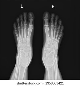
Normal Foot Radiology Case Radiopaedia Org
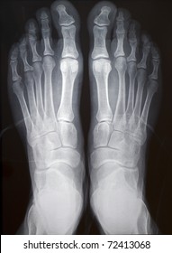
Foot Xray Images Stock Photos Vectors Shutterstock
1

Normal Ap Radiograph Of The Right Foot It Demonstrates The Larger Size Download Scientific Diagram
Left Foot Xray Images Stock Photos Vectors Shutterstock
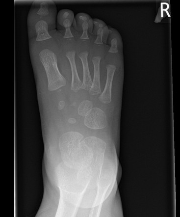
Foot X Ray Normal 2 Year Old Radiology Case Radiopaedia Org
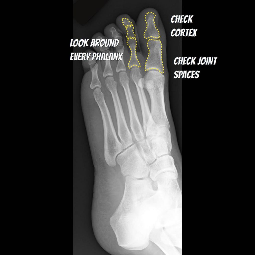
Foot Annotated X Ray Radiology Case Radiopaedia Org


